
The tarsal bones are found beneath and in front of the ankle. These are small bones that make the foot and ankle flexible, and help act as shock absorbers during walking and running. These bones include the calcaneus (heel bone), talus, navicular, cuboid and cuneiform.
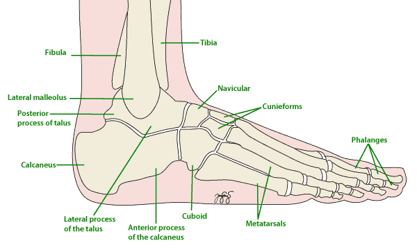
Bones of the Foot and Ankle
Symptoms
Many people with a tarsal coalition have no symptoms at all and never know they have one. Where symptoms do occur, they usually begin in the early teens, as the bones mature. The signs and symptoms include:
- Pain in the foot or ankle, often deep and hard to point to
- Pain that is worse with activity and eases with rest
- A flat foot that does not change shape when standing on tiptoe
- Stiffness in the foot, particularly turning the sole inwards and outwards
- Repeated ankle sprains, or a feeling that the ankle gives way
- Muscle spasm along the outside of the leg, which holds the foot turned outwards
Causes
A tarsal coalition is present from birth. During development, two of the tarsal bones fail to separate properly and remain joined by bone, cartilage or fibrous tissue.
The join limits the normal movement between the bones. Because the foot cannot absorb shock and adapt to the ground as it should, the surrounding joints take more load. This is what causes pain, and over time it can lead to arthritis in the neighbouring joints.
Symptoms often appear in adolescence because a coalition made of cartilage gradually turns to bone as the skeleton matures, making the foot progressively stiffer.
Risk Factors
Tarsal coalition often runs in families. Factors associated with symptoms include:
- A family history of tarsal coalition
- Adolescence, when the coalition stiffens as the skeleton matures
- Sport and activity that load the foot heavily
- Repeated ankle sprains, which are often the first sign
Investigations
Your doctor will examine the foot, looking at its shape when standing and checking how much the foot turns in and out. Tests that may be used include:
- X-ray, which often shows the coalition and any changes in the nearby joints
- CT scan, which shows a bony coalition best and is used when surgery is being planned
- MRI, which is better for a coalition made of cartilage or fibrous tissue
Complications
Problems that can follow an untreated coalition include:
- Pain that persists and limits walking and sport
- Arthritis in the joints around the coalition
- Repeated ankle sprains from a stiff foot that cannot adapt to uneven ground
- A flat foot that becomes fixed rather than flexible
Treatment
Many people need no treatment at all. Where the foot is painful, non-surgical treatment is tried first:
- Reducing activity for a period to let the symptoms settle
- Orthotics or supportive shoes to reduce movement at the painful joints
- Simple pain relief and anti-inflammatory medication
- A period in a boot or cast for a bad flare
- A steroid injection, in selected cases
Where pain persists despite these measures, surgery may be considered. In younger people with no arthritis, the coalition can be removed and the gap filled with fat or muscle to stop it re-forming. Where the surrounding joints are already worn, fusing the affected joints gives more reliable pain relief.
Prevention
A tarsal coalition is present from birth and cannot be prevented. What can be influenced is the pain it causes, by keeping to supportive footwear, managing activity sensibly, and having a stiff or repeatedly sprained foot assessed rather than accepting it.
F.A.Q. | Frequently Asked Questions
What is a tarsal coalition?
It is an abnormal join between two bones in the back of the foot that should be separate. The join is present from birth but usually only causes trouble in the early teens, when it hardens into bone and the foot becomes stiff and painful.
Why did it only start hurting now?
In childhood the join is soft cartilage and the foot still moves. As the child grows the join turns to bone, the foot loses its side to side movement and the joints around it are strained. Symptoms often begin after a sprain or a growth spurt.
Can it be treated without surgery?
Yes, at first. A period in a cast or boot, then a supportive insole and a change of activity, settles symptoms in many teenagers. If pain keeps returning, surgery to remove the join is considered.
What does surgery involve?
The bony bridge is cut out and fat or tendon is placed in the gap to stop it re forming. If the joints around the coalition are already arthritic, fusing them may be a better option.