

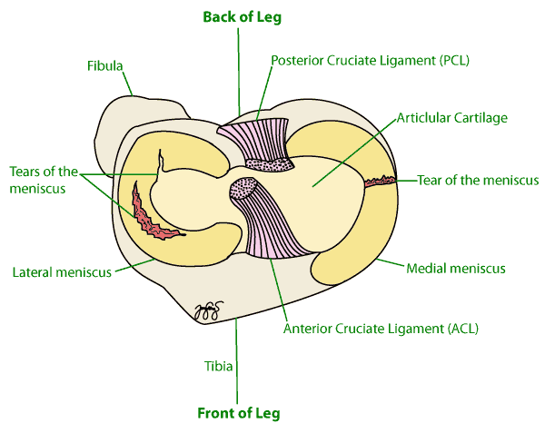
The meniscus is a structure made up of cartilage that is located in your knee joint. In each knee, there are two C-shaped menisci. They have the important function of acting as shock absorbers in your knee.
Your meniscus can be torn with a twisting injury to the knee. In older adults, the meniscus can be injured through a wear and tear process.
Symptoms
Some symptoms you may experience include:
- Pain immediately or when moving your knee
- Your knee may feel unstable
- Swelling which may worsen after the insult to your knee
- Stiffness of the knee
- May feel like your knee is locked or you may even feel a popping sensation
You may also have pain when walking or bearing weight.
Causes
The cause of a torn meniscus can be from any activity that causes you to forcefully twist your knee. This can be from sports or even when doing everyday activities like kneeling, squatting or heavy lifting.
In athletes, a torn meniscus is often associated with injuries to ligaments in your knee.
Risk Factors
Sports or activities in which there is twisting and pivoting of the knee puts the meniscus at risk of tearing. The sports in which tear is particularly common are contact sports, such as martial arts or football. Also activities involving a lot of pivoting like tennis or basketball have an increased risk of meniscal tear. As you get older, the risk of a torn meniscus increases because of the years of wear and tear on your knees.
Investigations
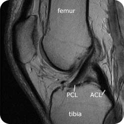
An MRI showing normal knee anatomy and an intact ACL

An MRI showing a tear of the meniscus
Clinical Examination: Often, a torn meniscus can be identified during a physical exam. Your doctor may bend your knee and perform physical exam manoeuvres to detect a meniscal tear.
X-rays: Your doctor may also obtain an X-ray of the knee to look for signs of osteoarthritis.
MRI: Magnetic resonance imaging (MRI) is a technique that uses a magnetic field and radio waves to create cross-sectional images of your knee. This is to confirm the diagnosis or study the extent of the tear.
Complications
The meniscus is a very important structure. It acts as a shock-absorber which reduces the pressure that is transmitted through your knee.
Your knee will wear out quicker if your meniscus is injured because more pressure is being put through your knee. This increased pressure can eventually lead to increased wearing out of your cartilage and osteoarthritis.
The injured meniscus can flip in your knee joint, causing a clicking sensation when the knee is bent. It can also lead to locking of your knee. Also, a torn meniscus can result in persistent knee pain.
Treatment
Treatment for a torn meniscus often begins conservatively. Your doctor may recommend:
- Rest: This is quite obvious, by resting and avoiding activities that aggravate your knee pain, you are preventing the injury from getting worse. Crutches are a good idea to take pressure off your knee and help your knee heal better.
- Ice: Ice can reduce knee pain and swelling and you can use an ice-pack or even just a bag of frozen vegetables.
- Compression: This can reduce the swelling in the knee
- Elevation: This also can reduce the level of swelling
- Medication: Over-the-counter pain relievers also can help ease knee pain.
- Knee exercises: Physiotherapy and rehabilitation can help you strengthen and stabilise the muscles around your knee and in your legs.
In middle-aged people, worn meniscus tears that do not cause locking do as well with physiotherapy as with keyhole surgery.
Surgery
If all the above conservative treatments fail and your knee remains painful, stiff or locked, surgery may be recommended. Sometimes the meniscus is repaired and other times it is trimmed. In both cases the approach is arthroscopic.
This arthroscopic surgery involves your doctor inserting an instrument called an arthroscope through a small incision near your knee. This equipment contains a light and a small camera, which shows the inside of your knee magnified onto a screen.
Recovery time following arthroscopic surgery tends to be much faster than it is for open-knee procedures because only small incisions are made. You may even be able to go home the same day. Nevertheless, full recovery may take weeks or months.
If the meniscus has degenerated and worn away, as would occur with severe degenerative arthritis, your doctor may recommend a knee joint replacement.
When a torn meniscus can be stitched, repair is preferred, as removing part of it raises the chance of arthritis and of needing a knee replacement later.
Seeking Advice
Your Family Doctor (GP)
Your Family Doctor will be able to diagnose and help treat your problem. He or she will be able to
- tell you about your problem
- advise you of the best treatment methods
- prescribe you medications
- and if necessary, refer you to Specialists (Consultants) for further treatment
F.A.Q. | Frequently Asked Questions
Can a meniscal tear heal by itself?
As it does not have its own blood supply, it can’t always get the required nutrients for self-repair. Thus it depends on where it is torn, but generally menisci have very poor ability to heal by themselves.
What is a meniscectomy?
It is a fairly simple operation in which all or part of the torn meniscus is removed.
Can an X-ray pick up a meniscal tear?
No, an X-ray is performed to rule out the presence of arthritis or bony fragments. X-rays look at bones while soft tissue (such as your meniscus) can be picked up via MRI.
After the operation when can I expect to notice a decrease in my pain?
Most patients notice a decreased pain after about a week. However, if there is pre-existing arthritis it may take 6 weeks to notice a difference.
When can I return to sport?
Usually in around 4–6 weeks. However, if you have a meniscal repair it will take longer, usually around 4 to 6 months.
When can I fly after my operation?
It is recommended not to fly on an aeroplane for a week after surgery due to the concern of leg swelling and blood clots called Deep Vein Thrombosis.
References
- D’Ambrosi R, Meena A, Raj A, Ursino N, Mangiavini L, Herbort M, Fink C. In elite athletes with meniscal injuries, always repair the lateral, think about the medial! A systematic review. Knee Surg Sports Traumatol Arthrosc. 2023, 31(6), 2500-2510. PMID 36319751.
- Migliorini F, Pilone M, Bell A, Celik M, Konrads C, Maffulli N. Outside-in repair technique is effective in traumatic tears of the meniscus in active adults. A systematic review. Knee Surg Sports Traumatol Arthrosc. 2023, 31(10), 4257-4264. PMID 37314454.
- Migliorini F, Oliva F, Eschweiler J, Cuozzo F, Hildebrand F, Maffulli N. No evidence in support of arthroscopic partial meniscectomy in adults with degenerative and nonobstructive meniscal symptoms. A level I evidence-based systematic review. Knee Surg Sports Traumatol Arthrosc. 2023, 31(5), 1733-1743. PMID 35776158.
- Migliorini F, Schäfer L, Bell A, Weber CD, Vecchio G, Maffulli N. Meniscectomy is associated with a higher rate of osteoarthritis compared to meniscal repair following acute tears. A meta-analysis. Knee Surg Sports Traumatol Arthrosc. 2023, 31(12), 5485-5495. PMID 37812251.